By Daebbang
“Since my mid-30s, brown patches have been spreading over my cheekbones. I was told it is melasma. Will it go away if I get laser treatment? I also heard there is a pill for it.”
It is right to think of melasma not as a blemish that can be removed in one go, but as a chronic pigment disorder that you lighten and then keep from darkening again. In skin with melasma, there is not only more pigment in the epidermis; changes in the dermis are also commonly seen together, such as damage to the boundary membrane between the epidermis and dermis, sun-damaged fibers and increased blood vessels[1, 2]. So in my judgment, melasma should be seen as a state in which the dermal environment has broken down, and restoring the dermal environment should also be a treatment goal alongside treatments that remove pigment.
The foundation of treatment is daily sun protection and topical lightening treatment. The international expert consensus is that other topical medicines such as azelaic acid and kojic acid, as well as oral tranexamic acid, are used as alternatives, that peels and microneedling are used as adjunctive treatments that support topical treatment, and that lasers are chosen carefully when other treatments are not enough[3]. Even after treatment lightens melasma, it commonly darkens again[3, 4].
There are also several pigment disorders that look like melasma but have different causes and treatments[5]. So melasma treatment starts with telling whether your patch is really melasma.
In this article, I will explain, in order, what melasma is and why it develops, how to tell melasma apart from look-alike pigment disorders by their borders and shape, how effective sun protection, topical medicines, oral medicine and peels are, and how far the evidence goes for the lasers many people get these days and for procedures that restore the dermal environment.
What is melasma?
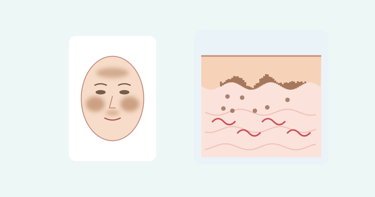
Melasma is a light brown to dark brown patch that appears symmetrically on both sides of the face. It is characterized by irregular borders and a shape that spreads widely like a net. It is most common in the central face, such as the forehead, nose, upper lip and cheeks, and it can also appear only on the cheekbones or along the jawline. It is more common in women and in people with darker skin. How common it is varies widely, from 1% to 50%, depending on the population studied[6].
Under the microscope, melanin pigment is increased in the epidermis, the outer layer of the skin. In a study that compared the skin of 56 Korean patients with melasma with the normal skin right next to it, the melasma areas had more melanin in every layer of the epidermis, and the pigment-producing cells were also more active. Signs of sun damage were also more pronounced[7]. In some cases, there is also pigment in the dermis, the inner layer of the skin, or the small blood vessels are increased[6, 8].
Looking more deeply, melasma also involves changes in the dermis. Between the epidermis and dermis there is a thin boundary membrane called the basement membrane. In a study that examined the skin of 48 patients with melasma, depending on the staining method, this basement membrane was damaged in 83–95.5% of the tissue from melasma areas. Pigmented basal cells of the epidermis were often seen hanging down toward the dermis, and mast cells were increased in the dermis[9]. In a study that looked at the skin of 10 women with melasma under an electron microscope, the basement membrane in melasma areas was also broken or thinned in places. The spots where pigment cells sagged down toward the dermis were where the basement membrane was broken[10].
The dermis itself is also changed. In 83–93% of patients with melasma, solar elastosis, in which sun-damaged elastic fibers are clumped together, was seen[2]. A review that summarized dermal changes in melasma said that basement membrane damage, sagging pigment cells, solar elastosis, cells that have engulfed pigment, mast cells and new blood vessels are commonly seen in melasma, and that these dermal changes may be connected with the pigment changes in the epidermis. It also considered that these changes could become new targets for treatment[1]. There is also a view that melasma should be regarded as a skin disorder caused by sun-induced aging[11].
In my judgment, the fact that melasma spreads widely without clear borders and keeps darkening again even after treatment is closely related to this broken-down dermal environment. This is why treatment that only removes pigment from the epidermis is not enough and the dermal environment must be restored as well.


Why does melasma develop?
Sunlight, female hormones and family history are considered the main causes[6]. One study compared 207 Brazilian women with melasma and 207 women of the same age without melasma. The proportion who said a family member had melasma was 61% in the melasma group and 13% in the group without it. A lifestyle with long sun exposure, a history of pregnancy, the duration of oral contraceptive use and irregular periods were also associated with melasma[13].
Not only ultraviolet light but also visible light, the light we can see, is involved. Visible light can cause pigmentation in people with darker skin[14]. Because small blood vessels are often increased in melasma areas, changes in blood vessels are also being studied as one of the causes[8].
In my judgment, this is what makes melasma most different from other blemishes. As long as there are day-to-day triggers such as sunlight and hormones, melasma can darken again. That is why the word “management” suits it better than “treatment.”
What pigment disorders are confused with melasma?
Brown or gray patches on the face are divided into epidermal, dermal and mixed types according to the depth in the skin at which the pigment is located[5]. Pigment disorders that need to be distinguished from melasma include freckles, lentigines, post-inflammatory hyperpigmentation, acquired bilateral nevus of Ota-like macules and nevus of Ota[6]. When the depth differs, the treatment that works well also differs.
The first thing to look at with the eye is the border. Melasma is a patch with irregular borders that spreads widely like a net[6]. Some melasma can also look sharply bordered[15]. Freckles are small light brown dots of 1–2 mm that appear one by one, separately; they start in childhood and increase in number at puberty. Lentigines are flat brown spots that form on sun-exposed areas, with sharp borders[16]. However, facial pigment disorders often overlap in their appearance and dermoscopic findings, so the diagnosis should not be settled by the border alone; the history, examination and dermoscopy (a magnifying skin examination) should be considered together[17].

| Pigment disorder | Shape and location | Color and depth | Treatment approach |
|---|---|---|---|
| Melasma | Patches with irregular borders that spread widely like a net, symmetric on the forehead, nose, upper lip, cheeks, cheekbones and jawline[6] | Light brown to dark brown, mainly epidermis (partly dermis) | Sun protection and topical medicines as the foundation, lasers with caution[3, 6] |
| Freckles | Small 1–2 mm dots scattered separately one by one, start in childhood and increase at puberty[16], clear genetic influence[19] | Brown, epidermis | Need to be distinguished from melasma[6] |
| Lentigines (solar lentigines) | Flat brown spots with sharp borders[16], on areas exposed to the sun for a long time, an early sign of skin aging | Brown, epidermis | Laser, cryotherapy and others[20] |
| Post-inflammatory hyperpigmentation | Where there was inflammation, such as acne, wounds or procedures | Varies with the type and depth of inflammation | Needs to be distinguished from melasma, covered in detail in the article below[6] |
| Acquired bilateral nevus of Ota-like macules | Small dots on both sides of the face in middle-aged women, especially Asian women | Gray-brown to gray-blue, dermis | Pigment-targeting laser is the main treatment[12] |
| Nevus of Ota | Mainly Asian children and women, around one eye and on the forehead and cheek | Gray-blue, dermis | Pigment-targeting laser (Q-switched laser)[18] |
In particular, acquired bilateral nevus of Ota-like macules is a dermal pigment disorder common in Asian women, and because it appears on the face, it is easily confused with melasma. Because the pigment sits at the middle depth of the dermis, unlike melasma, a laser that targets pigment is the main treatment[12]. In a study that compared the skin of 50 people with melasma and 46 people with these macules with dermoscopy, melasma was mostly light brown, while these macules were more often gray or blue-brown. Even when the two were present together, dermoscopy helped tell them apart[21]. Before getting laser treatment, it is good to confirm in a clinic visit what kind of pigment it is. I covered the difference between freckles and melasma and the difference between pico toning and spot treatment in Why Are My Freckles and Dark Spots Still There After Pico Toning?, and post-inflammatory hyperpigmentation in detail in Dark Patches After Laser Treatment: Why Does Post-Inflammatory Hyperpigmentation Happen, and How Long Does It Last?.
Why is sun protection the most important?
The international expert consensus named daily use of a broad-spectrum sunscreen as the foundation for every patient with melasma[3]. However, ordinary sunscreens do not block visible light well enough. To block visible light as well, a tinted sunscreen containing pigments such as iron oxide, which leave color on the skin, is helpful[14].
In one randomized study, all 68 patients with melasma applied 4% hydroquinone and were divided into a group using a sunscreen that blocks only ultraviolet light and a group using an iron oxide sunscreen that also blocks visible light. After 8 weeks, the group that also blocked visible light improved 15% more in melasma score[22]. There are also randomized studies in which melasma recurred less in the group that used a sunscreen with added visible-light-blocking ingredients[6].
In my judgment, if you have melasma, it is good to choose a tinted sunscreen and to apply it even indoors if you spend a long time near a window. Use physical protection such as hats and parasols as well.
What topical medicines are there?
Topical medicines are still the center of melasma treatment. An evidence-based review that summarized 113 studies (6,897 people) concluded that hydroquinone alone and triple combination cream (a cream that combines hydroquinone, tretinoin and a steroid) are the most effective and most studied treatments[23]. In a Cochrane review that examined 20 randomized studies (2,125 people), triple combination cream also lightened melasma more than hydroquinone alone. 20% azelaic acid was more effective than 2% hydroquinone, but no clear difference from 4% hydroquinone was found[24].
These medicines should be used under a doctor’s prescription and supervision. The international expert consensus also named triple combination cream as standard treatment while saying that it should be adjusted and used under a doctor’s supervision[3]. Applying hydroquinone for a long time can cause exogenous ochronosis, in which the skin instead turns blue-black, and once it develops, it is very difficult to treat[25]. Long-term use of the steroid in triple combination cream can thin the skin or make small blood vessels visible[6]. When hydroquinone is hard to use, other topical medicines such as azelaic acid or kojic acid are used[3].
Does oral tranexamic acid work?
Tranexamic acid was originally a medicine that reduces bleeding. In melasma, it is being studied as a treatment that suppresses the process by which pigment is made and also acts on the blood vessel changes of melasma[6, 8]. In one randomized study, 44 people with moderate to severe melasma took tranexamic acid or a placebo for 3 months, and all of them used sunscreen. After 3 months, the melasma score decreased by 49% in the tranexamic acid group and by 18% in the placebo group. However, 3 months after stopping the medicine, the difference had narrowed greatly, to 26% and 19%[26]. In an analysis that pooled 24 randomized studies of tranexamic acid used by mouth, on the skin or as intradermal injection, tranexamic acid also increased the effect when added to existing treatment[27].
Because tranexamic acid is a medicine that reduces bleeding, blood clots are the biggest concern. In a summary that pooled randomized studies of oral tranexamic acid, no blood clot events were observed[28]. However, long-term safety needs more research[23]. A consensus of pigment disorder experts also said that a history of blood clots, cardiovascular disease and menstrual history should be checked before starting oral tranexamic acid[29]. Before taking oral tranexamic acid, you must see a doctor and tell them about your own medical history, such as whether you have ever had a blood clot, and the medicines you are taking. Because melasma tends to darken again when the medicine is stopped, topical treatment and sun protection should be continued while you take it.
Do peels help?
A chemical peel is a treatment that uses a mild acid to thinly remove the outer layer of the skin and help pigment come out. In a systematic review that summarized 24 studies (1,075 people) of peels for melasma, peels were safe and effective, and glycolic acid peels were the safest and most effective. However, because the methods differed from study to study, the results could not be combined into one analysis[30]. The international expert consensus recommended peels as an adjunctive treatment that increases the effect of topical treatment[3].
Peels also create inflammation in the skin, so if they are done too strongly, post-inflammatory hyperpigmentation can develop. In my judgment, the safe order is to start with topical treatment first and then add a mild peel on top of it.
What about the lasers and procedures people get these days?
Laser toning is a method in which a 1064 nm Q-switched Nd:YAG laser is applied over the whole face at low fluence in multiple passes. In an analysis that pooled 50 studies (1,772 people), toning alone lightened melasma at first, but its effect decreased over time, and after 24 weeks there was almost no difference from before treatment. Combining it with topical medicines and other treatments was more effective than toning alone. Side effects included hypopigmentation, in which color is lost and the skin turns white, post-inflammatory hyperpigmentation and recurrence, and hypopigmentation was more common with more treatment sessions[31]. A systematic review of 42 studies by Korean researchers also said that, although rare, mottled hypopigmentation can develop and last a long time, and that doing it too strongly can instead cause pigmentation[32].
A picosecond laser is a method that fires the laser in very short bursts. In an analysis that pooled 6 randomized studies, the 1064 nm picosecond laser reduced the melasma score, but the 755 nm picosecond laser did not come out better than topical lightening agents and caused post-inflammatory hyperpigmentation[33]. Even with the same picosecond laser, the results differ with wavelength and intensity.
Results for fractional lasers are mixed. In a randomized study in which one side of the face of 29 people was treated with a non-ablative fractional laser and the other side with triple combination cream, the laser side instead became darker, and 9 people (31%) developed post-inflammatory hyperpigmentation[34]. In an analysis that pooled 22 laser studies (694 people), several lasers reduced the melasma score, but people with darker skin had a risk of post-inflammatory hyperpigmentation and hypopigmentation[35].
Microneedling has evidence when used together with topical medicines. In an analysis that pooled 12 studies (459 people), adding microneedling to topical medicines led to greater improvement at 12–16 weeks than topical medicines alone, and no serious side effects were reported[36]. In an analysis that compared 59 randomized studies together, Q-switched Nd:YAG laser, ablative fractional laser, triple combination cream and oral tranexamic acid ranked ahead, and in 27 of the 31 studies that made the comparison, combined treatment was better than a single treatment[37].
In my judgment, lasers can help lighten melasma, but on their own they are not a treatment that ends melasma. The international expert consensus also said to use lasers when melasma does not respond well to other treatments[3]. If you have had dozens of toning sessions at short intervals and mottled white spots have appeared or the area has instead darkened, stop rather than adding more sessions and see a doctor again. I covered how pico toning differs between melasma and other blemishes in Why Are My Freckles and Dark Spots Still There After Pico Toning?.
Do treatments that restore the dermal environment work?
As we saw above, skin with melasma commonly also has dermal changes. There is a proposal to make these dermal changes a treatment target[1], and besides treatments that break up pigment directly, treatments that aim to restore the dermis and reduce oxidative stress (damage caused by reactive oxygen) and inflammation are being studied as new approaches[38, 39, 40]. Representative ones are low-fluence long-pulsed Nd:YAG laser toning, PDRN, PN and mixed cross-linked and non-cross-linked hyaluronic acid injections (skin boosters).
Low-fluence long-pulsed Nd:YAG laser toning (commonly called “Genesis toning”) is a method that fires the same 1064 nm Nd:YAG laser in microsecond-range pulses, much longer than Q-switched pulses. In one study, the laser was applied until the skin temperature rose to about 40–42°C and the skin turned evenly and slightly red[41]. In a study in which 30 patients whose melasma had instead darkened after other treatments received Q-switched toning combined with long-pulsed toning 10 times, the melasma score decreased from 10.48 to 3.22. The researchers interpreted that the long-pulsed laser may stabilize melasma by remodeling the dermis[42]. In a study of 21 people in which one side of the face received Q-switched toning only and the other side received added microsecond toning, the side with the added treatment had lower melanin values even at 12 months, and redness also decreased more. However, after 6 months, the melasma score showed no clear difference from before treatment[43]. In a randomized study that divided 40 people, the melasma score decreased both in the group that used Q-switched and long-pulsed lasers together and in the picosecond alexandrite laser group, and it decreased more in the combined group. Recurrence at 24 weeks was 0% and 10.5%, with no statistically significant difference[44]. This study did not look separately at the effect of adding the long-pulsed laser; it compared two laser approaches.
PDRN (polydeoxyribonucleotide) is an ingredient made from DNA fragments that has been used to help wound healing and collagen production in sun-aged skin. In cell experiments, PDRN reduced melanin and the enzyme that makes pigment, and there is also an observation that facial hyperpigmentation visibly lightened when 6 women with it received 3 injections into the skin[38]. Another cell experiment also showed an antioxidant effect that removes reactive oxygen and an effect that reduces the enzyme that breaks down collagen[45].
PN (polynucleotide) is a DNA-fragment ingredient with a structure similar to PDRN (for example, Rejuran). In experiments with cells and animal skin exposed to ultraviolet light, PN reduced oxidative stress and inflammatory signals and also reduced melanin buildup[39]. However, in a randomized study in which 30 patients with melasma received radiofrequency microneedling on both sides of the face and then had PN applied to only one side, there was no difference between the two sides up to 6 months[46].
Mixed cross-linked and non-cross-linked hyaluronic acid injection is a skin booster that mixes hyaluronic acid whose molecules are partly linked together (cross-linked) with hyaluronic acid that is not linked (non-cross-linked) (for example, HILOWAVE). In experiments with cells and rats, a hybrid hyaluronic acid in which hyaluronic acids of different molecular sizes were lightly linked increased collagen and elastic fiber production, showed antioxidant and anti-inflammatory effects, suppressed genes that make pigment and reduced melanin built up by ultraviolet light[40]. Human melasma studies are small studies with similar injections. In 6 people whose toning results had stopped improving, melasma lightened further when toning was continued after injection of non-cross-linked hyaluronic acid containing antioxidant ingredients[47]. In a randomized study (36 of 50 people completed it) that added a hyaluronic acid skin booster containing glutathione to Q-switched toning and oral tranexamic acid treatment, the melasma score of the group with the added treatment was 2.50 at 6 months, lower than 3.39 in the group without it, and the difference was still maintained 2 months after treatment[48]. Because this study also included an antioxidant ingredient called glutathione, it is hard to regard the result as the effect of hyaluronic acid alone.
| Treatment | Expected action | Level of evidence in melasma |
|---|---|---|
| Low-fluence long-pulsed Nd:YAG laser toning | Gently warms the dermis to restore it | Small studies used together with Q-switched toning. In a split-face study that looked at the added effect, the melasma score showed no difference after 6 months[42, 43, 44] |
| PDRN | Suppresses pigment production, antioxidant, suppresses collagen-degrading enzyme | Cell experiments and an observation of 6 people with facial hyperpigmentation (not limited to melasma)[38, 45] |
| PN | Antioxidant, suppresses inflammatory signals, reduces melanin | Cell and animal experiments. No added effect in a randomized study that added it to radiofrequency microneedling[39, 46] |
| Mixed cross-linked and non-cross-linked hyaluronic acid injection | Collagen and elastic fiber production, antioxidant, anti-inflammatory | Cell and animal experiments. An observation of 6 people and 1 randomized study with similar hyaluronic acid injections[40, 47, 48] |
In my judgment, treatments that restore the dermal environment are meaningful in that they target the reason melasma spreads widely and keeps recurring. However, there are still few studies in people, and most are test-tube and animal experiments or small clinical studies. So they are not treatments that replace sun protection and topical medicines; it is right to use them together as adjunctive treatments added to the basic treatment.
How can I keep it from darkening again?
Even after melasma has lightened with treatment, it commonly darkens again. The international expert consensus also named the high recurrence rate as a reason melasma is hard to manage and said that long-term maintenance strategies need more research[3]. A Korean review explains that recurrence is frequent because aging changes such as sun damage and increased blood vessels are also present in skin with melasma[4]. In a randomized study in which people whose melasma had cleared or become mild after 8 weeks of triple combination cream received maintenance treatment for 6 months by applying it less often, 53% stayed free of recurrence[49].
In my judgment, sun protection must continue even after melasma has lightened. Rather than stopping topical medicines right away, it is also good to decide with your doctor whether to continue them less often. Knowing in advance about situations related to melasma, such as a lifestyle with a lot of sun exposure, pregnancy and oral contraceptives, and preparing for them also helps[13].
So what should I do?
| Situation | My opinion | Reason |
|---|---|---|
| When you are not sure whether the patch is melasma | Before any procedure, first confirm the type of pigment disorder at a dermatology visit | An irregular patch that spreads widely suggests melasma first and a sharply bordered spot suggests a lentigo first, but appearances overlap, so examination and dermoscopy are needed[6, 16, 17] |
| When treating melasma for the first time | Apply a tinted sunscreen every day and start with prescription topical medicine | International consensus named sun protection and triple combination cream as the foundation[3, 22] |
| When topical medicine is not enough | Discuss adding oral tranexamic acid, a mild peel or microneedling | Adding them to existing treatment increased the effect[3, 27, 36] |
| When considering oral tranexamic acid | Be sure to see a doctor before taking it and tell them your medical history and the medicines you take | Blood clot events were not reported, but long-term safety needs more research. Expert consensus also recommended checking medical history before taking it[23, 28, 29] |
| When you are getting laser toning | Do not keep adding sessions without limit, and stop if white spots or darkening appear | Hypopigmentation was more common with more sessions, and the effect decreased over time[31, 32] |
| When melasma spreads widely and keeps recurring | Discuss adding treatments that restore the dermal environment to the basic treatment | Dermal changes are common in skin with melasma, but for dermal treatments, small studies include both results in which they helped and results that showed no difference[1, 43, 46, 48] |
| After melasma has lightened | Continue sun protection and decide on maintenance treatment with your doctor | Even with maintenance treatment, only about half stayed free of recurrence for 6 months[49] |
Take-home messages
- Melasma is a brown patch that appears symmetrically on both sides of the face, and sunlight, female hormones and family history are the main causes. Not only pigment in the epidermis but also dermal changes such as basement membrane damage, sun-damaged fibers and increased blood vessels are commonly present.
- Melasma is a patch with irregular borders that spreads widely, freckles are small dots scattered separately, and lentigines are sharply bordered spots. Pigment disorders that look like melasma differ in depth and treatment, so first confirm the type with an examination before treatment.
- Daily sun protection is the foundation of treatment. A tinted sunscreen that also blocks visible light helps more.
- Prescription topical medicines, especially triple combination cream, are the treatment with the most evidence. However, they should be used under a doctor’s supervision.
- Oral tranexamic acid increases the effect of existing treatment, but melasma tends to darken again when it is stopped. Be sure to see a doctor before taking it.
- Laser toning can help, but its effect on its own decreases over time, and getting it too often can cause white spots. Results with picosecond lasers differed by wavelength.
- Treatments that restore the dermal environment, such as long-pulsed Nd:YAG laser toning, PDRN, PN and hyaluronic acid skin boosters, still have few studies, so they are used together as adjunctive treatments added to the basic treatment.
- Melasma commonly recurs even after it has lightened, so sun protection and maintenance treatment must continue.
References
[1] Phansuk K, Vachiramon V, Jurairattanaporn N, et al. Dermal Pathology in Melasma: An Update Review. Clinical, Cosmetic and Investigational Dermatology. 2022;15:11–19. DOI: 10.2147/CCID.S343332
[2] Kwon SH, Hwang YJ, Lee SK, et al. Heterogeneous Pathology of Melasma and Its Clinical Implications. International Journal of Molecular Sciences. 2016;17(6):824. DOI: 10.3390/ijms17060824
[3] Sarkar R, Desai SR, Sinha S, et al. Delphi consensus on melasma management by international experts and pigmentary disorders society. Journal of the European Academy of Dermatology and Venereology. 2026;40(4):680–692. DOI: 10.1111/jdv.70066 (research funding from a pharmaceutical company)
[4] Jo JY, Chae SJ, Ryu HJ. Update on Melasma Treatments. Annals of Dermatology. 2024;36(3):125–134. DOI: 10.5021/ad.23.133
[5] Wang RF, Ko D, Friedman BJ, et al. Disorders of hyperpigmentation. Part I. Pathogenesis and clinical features of common pigmentary disorders. Journal of the American Academy of Dermatology. 2023;88(2):271–288. DOI: 10.1016/j.jaad.2022.01.051 (some authors participated in research for or advised cosmetic and pharmaceutical companies)
[6] Ogbechie-Godec OA, Elbuluk N. Melasma: an Up-to-Date Comprehensive Review. Dermatology and Therapy. 2017;7(3):305–318. DOI: 10.1007/s13555-017-0194-1
[7] Kang WH, Yoon KH, Lee ES, et al. Melasma: histopathological characteristics in 56 Korean patients. British Journal of Dermatology. 2002;146(2):228–237. DOI: 10.1046/j.0007-0963.2001.04556.x
[8] Masub N, Nguyen JK, Austin E, et al. The Vascular Component of Melasma: A Systematic Review of Laboratory, Diagnostic, and Therapeutic Evidence. Dermatologic Surgery. 2020;46(12):1642–1650. DOI: 10.1097/DSS.0000000000002770
[9] Torres-Álvarez B, Mesa-Garza IG, Castanedo-Cázares JP, et al. Histochemical and immunohistochemical study in melasma: evidence of damage in the basal membrane. American Journal of Dermatopathology. 2011;33(3):291–295. DOI: 10.1097/DAD.0b013e3181ef2d45
[10] Espósito ACC, Brianezi G, de Souza NP, et al. Ultrastructural characterization of damage in the basement membrane of facial melasma. Archives of Dermatological Research. 2020;312(3):223–227. DOI: 10.1007/s00403-019-01979-w
[11] Passeron T, Picardo M. Melasma, a photoaging disorder. Pigment Cell & Melanoma Research. 2018;31(4):461–465. DOI: 10.1111/pcmr.12684
[12] Park JM, Tsao H, Tsao S. Acquired bilateral nevus of Ota-like macules (Hori nevus): etiologic and therapeutic considerations. Journal of the American Academy of Dermatology. 2009;61(1):88–93. DOI: 10.1016/j.jaad.2008.10.054
[13] Handel AC, Lima PB, Tonolli VM, et al. Risk factors for facial melasma in women: a case-control study. British Journal of Dermatology. 2014;171(3):588–594. DOI: 10.1111/bjd.13059
[14] Lyons AB, Trullas C, Kohli I, et al. Photoprotection beyond ultraviolet radiation: A review of tinted sunscreens. Journal of the American Academy of Dermatology. 2021;84(5):1393–1397. DOI: 10.1016/j.jaad.2020.04.079 (some authors affiliated with a sunscreen company)
[15] Bostan E, Cakir A. The dermoscopic characteristics of melasma in relation to different skin phototypes, distribution patterns and wood lamp findings: a cross-sectional study of 236 melasma lesions. Archives of Dermatological Research. 2023;315(7):1927–1938. DOI: 10.1007/s00403-023-02584-8
[16] Hua Y, Xie B, Shen Y, et al. Dermatoscopic Features of Hyperpigmented Dermatoses. Clinical, Cosmetic and Investigational Dermatology. 2026;19:609514. DOI: 10.2147/CCID.S609514
[17] Mansilla-Polo M, Díaz-Gómez CJ, Morgado-Carrasco D. Clinical and Dermoscopic Characteristics of Facial Hyperpigmentation Disorders in Skin of Color. Actas Dermo-Sifiliográficas. 2026;117(7):104637. DOI: 10.1016/j.ad.2026.104637
[18] Shah VV, Bray FN, Aldahan AS, et al. Lasers and nevus of Ota: a comprehensive review. Lasers in Medical Science. 2016;31(1):179–185. DOI: 10.1007/s10103-015-1834-2
[19] Luo S, Li Z, Wang M, et al. Genome wide association study and meta-analysis identified multiple new risk loci for freckles in 4813 Chinese individuals. Pigment Cell & Melanoma Research. 2024;37(6):808–821. DOI: 10.1111/pcmr.13183
[20] Mardani G, Nasiri MJ, Namazi N, et al. Treatment of Solar Lentigines: A Systematic Review of Clinical Trials. Journal of Cosmetic Dermatology. 2025;24(4):e70133. DOI: 10.1111/jocd.70133
[21] Rattananukrom T, Suchonwanit P, Thadanipon K, Vachiramon V. A Comparative Study of Dermatoscopic Features of Melasma and Hori’s Nevus in Asian Patients. Journal of Clinical and Aesthetic Dermatology. 2022;15(3):16–20. PubMed: 35342501
[22] Castanedo-Cazares JP, Hernandez-Blanco D, Carlos-Ortega B, et al. Near-visible light and UV photoprotection in the treatment of melasma: a double-blind randomized trial. Photodermatology, Photoimmunology & Photomedicine. 2014;30(1):35–42. DOI: 10.1111/phpp.12086
[23] McKesey J, Tovar-Garza A, Pandya AG. Melasma Treatment: An Evidence-Based Review. American Journal of Clinical Dermatology. 2020;21(2):173–225. DOI: 10.1007/s40257-019-00488-w
[24] Rajaratnam R, Halpern J, Salim A, et al. Interventions for melasma. Cochrane Database of Systematic Reviews. 2010;(7):CD003583. DOI: 10.1002/14651858.CD003583.pub2
[25] Bhattar PA, Zawar VP, Godse KV, et al. Exogenous Ochronosis. Indian Journal of Dermatology. 2015;60(6):537–543. DOI: 10.4103/0019-5154.169122
[26] Del Rosario E, Florez-Pollack S, Zapata L Jr, et al. Randomized, placebo-controlled, double-blind study of oral tranexamic acid in the treatment of moderate-to-severe melasma. Journal of the American Academy of Dermatology. 2018;78(2):363–369. DOI: 10.1016/j.jaad.2017.09.053
[27] Feng X, Su H, Xie J. Efficacy and safety of tranexamic acid in the treatment of adult melasma: An updated meta-analysis of randomized controlled trials. Journal of Clinical Pharmacy and Therapeutics. 2021;46(5):1263–1273. DOI: 10.1111/jcpt.13430
[28] Kuceki G, Mendez D, Garza-Dueñas GG, et al. No thromboembolic events observed in randomized controlled trials of oral tranexamic acid: A systematic review and descriptive synthesis. Journal of the American Academy of Dermatology. 2025;93(4):1086–1088. DOI: 10.1016/j.jaad.2025.06.001
[29] Sarkar R, Narayan R V, Vinay K, et al. Prescribing practices of tranexamic acid for melasma: Delphi consensus from the Pigmentary Disorders Society. Indian Journal of Dermatology, Venereology and Leprology. 2023;90(1):41–45. DOI: 10.25259/IJDVL_1157_2022
[30] Sarkar R, Lakhani R. Chemical Peels for Melasma: A Systematic Review. Dermatologic Surgery. 2024;50(7):656–661. DOI: 10.1097/DSS.0000000000004167
[31] Cervantes J, Patzelt N, Al-Janahi S, et al. Efficacy and Safety of Low Fluence Nd:YAG Laser Treatment in Melasma: A Meta-Analysis and Systematic Review. Dermatologic Surgery. 2023;49(1):36–41. DOI: 10.1097/DSS.0000000000003635
[32] Lee YS, Lee YJ, Lee JM, et al. The Low-Fluence Q-Switched Nd:YAG Laser Treatment for Melasma: A Systematic Review. Medicina (Kaunas). 2022;58(7):936. DOI: 10.3390/medicina58070936
[33] Feng J, Shen S, Song X, et al. Efficacy and safety of picosecond laser for the treatment of melasma: a systematic review and meta-analysis. Lasers in Medical Science. 2023;38(1):84. DOI: 10.1007/s10103-023-03744-y
[34] Wind BS, Kroon MW, Meesters AA, et al. Non-ablative 1,550 nm fractional laser therapy versus triple topical therapy for the treatment of melasma: a randomized controlled split-face study. Lasers in Surgery and Medicine. 2010;42(7):607–612. DOI: 10.1002/lsm.20937
[35] Lai D, Zhou S, Cheng S, et al. Laser therapy in the treatment of melasma: a systematic review and meta-analysis. Lasers in Medical Science. 2022;37(4):2099–2110. DOI: 10.1007/s10103-022-03514-2
[36] Bailey AJM, Li HO, Tan MG, et al. Microneedling as an adjuvant to topical therapies for melasma: A systematic review and meta-analysis. Journal of the American Academy of Dermatology. 2022;86(4):797–810. DOI: 10.1016/j.jaad.2021.03.116
[37] Liu Y, Wu S, Wu H, et al. Comparison of the Efficacy of Melasma Treatments: A Network Meta-Analysis of Randomized Controlled Trials. Frontiers in Medicine. 2021;8:713554. DOI: 10.3389/fmed.2021.713554
[38] Noh TK, Chung BY, Kim SY, et al. Novel Anti-Melanogenesis Properties of Polydeoxyribonucleotide, a Popular Wound Healing Booster. International Journal of Molecular Sciences. 2016;17(9):1448. DOI: 10.3390/ijms17091448
[39] Oh S, Jeon HD, Rho NK, et al. Polynucleotide Mixture Attenuates Ultraviolet B-Induced Skin Pigmentation. International Journal of Molecular Sciences. 2025;26(13):6399. DOI: 10.3390/ijms26136399
[40] Roh H, Nguyen NH, Jung J, et al. Regenerative Skin Remodeling by a Dual Hyaluronic Acid Hybrid Complex in Multimodal Preclinical Models. International Journal of Molecular Sciences. 2026;27(2):1027. DOI: 10.3390/ijms27021027 (one author affiliated with a company research institute)
[41] Hai L, Phuong B, Ha L, et al. Dual Toning Method with the Combination of Picosecond and Microsecond Nd:YAG in Refractory Melasma Unresponsive to Picosecond Alone. Journal of Cutaneous and Aesthetic Surgery. 2021;14(1):101–106. DOI: 10.4103/JCAS.JCAS_30_20
[42] Choi CP, Yim SM, Seo SH, et al. Retreatment using a dual mode of low-fluence Q-switched and long-pulse Nd:YAG laser in patients with melasma aggravation after previous therapy. Journal of Cosmetic and Laser Therapy. 2015;17(3):129–134. DOI: 10.3109/14764172.2014.988730
[43] Wu X, Cen Q, Lin X, et al. Novel melasma therapy using combined low fluence and microsecond pulse Q switched 1064 nm neodymium doped yttrium aluminium garnet laser. Scientific Reports. 2025;15(1):24596. DOI: 10.1038/s41598-025-10129-4
[44] Liang S, Shang S, Tan A, et al. Comparative efficacy and safety of the novel Picosecond Alexandrite Laser and the traditional combined Q-switched and long-pulse Nd: YAG lasers in melasma treatment: a randomized evaluator-blinded trial. Lasers in Medical Science. 2025;40(1):29. DOI: 10.1007/s10103-025-04286-1
[45] Kim YJ, Kim MJ, Kweon DK, et al. Polydeoxyribonucleotide Activates Mitochondrial Biogenesis but Reduces MMP-1 Activity and Melanin Biosynthesis in Cultured Skin Cells. Applied Biochemistry and Biotechnology. 2020;191(2):540–554. DOI: 10.1007/s12010-019-03171-2 (one author affiliated with a company)
[46] Gulfan MCB, Wanitphakdeedecha R, Wongdama S, et al. Efficacy and Safety of Using Noninsulated Microneedle Radiofrequency Alone Versus in Combination with Polynucleotides for the Treatment of Melasma: A Pilot Study. Dermatology and Therapy. 2022;12(6):1325–1336. DOI: 10.1007/s13555-022-00728-8 (device and medications provided by companies)
[47] Yip DL. A pilot study on the use of a non-crosslinked hyaluronic acid with associated anti-oxidant ingredients to improve the effectiveness of Nd:YAG laser toning in the treatment of melasma in six patients. Journal of Cosmetic Dermatology. 2020;19(9):2212–2218. DOI: 10.1111/jocd.13565
[48] Bai JL. Hyaluronic acid-based skin booster with glutathione combined with 1064-nm Nd:YAG laser and oral tranexamic acid for melasma: A prospective comparative study. Medicine (Baltimore). 2026;105(42):e51212. DOI: 10.1097/MD.0000000000051212
[49] Arellano I, Cestari T, Ocampo-Candiani J, et al. Preventing melasma recurrence: prescribing a maintenance regimen with an effective triple combination cream based on long-standing clinical severity. Journal of the European Academy of Dermatology and Venereology. 2012;26(5):611–618. DOI: 10.1111/j.1468-3083.2011.04135.x


Leave a Reply