
By Daebbang
“I had a laser treatment to remove a spot, but the area where the scab fell off looks even darker. Did something go wrong? Will it stay like this?”
A dark patch after a procedure is most often first thought to be post-inflammatory hyperpigmentation (PIH). After the skin has been inflamed by heat or injury, the pigment melanin increases in that spot. It can follow skin conditions such as acne or eczema and burns, and it can also follow skin procedures such as lasers[1].
In short, it often fades with time, but how long that takes depends greatly on how deep the pigment sits. This article walks through why it happens, how long it lasts, and what to do and avoid in the meantime.
The laser removes pigment, so why can the skin get darker?
A laser delivers a set amount of energy to target pigment or tissue. In the process, the skin goes through a phase of small injury and recovery. Inflammatory signals during recovery stimulate the skin’s pigment cells, which then make more melanin and pass it on to neighboring skin cells[2].
An easy comparison is a little extra dust settling where construction has just finished. It does not mean the work was done wrong, but the area needs time to settle. The darker the skin tone, the more easily laser energy is also absorbed by pigment in the epidermis, so this reaction tends to be more common and more intense[1, 2].
PIH is also reported as a common side effect of laser treatment in Asian patients. A study from Hong Kong found that, even with the same laser, treatment settings such as energy and density, and whether the skin was cooled, may be related to the risk. However, it was a small retrospective study that looked back at past medical records, so it is not evidence that any particular setting is safe[3].
How long does it last? The ‘depth’ of the pigment decides
PIH is generally understood as two types, depending on how deep the pigment sits.
- Epidermal (shallow layer): Melanin has increased in the outermost layer of the skin. It often looks brown to dark brown.[2, 4]
- Dermal (deep layer): Inflammation damages the area below the epidermis, pigment drops into the dermis, and scavenger cells (macrophages) capture it and keep it there. It can look bluish-gray.[2, 4]
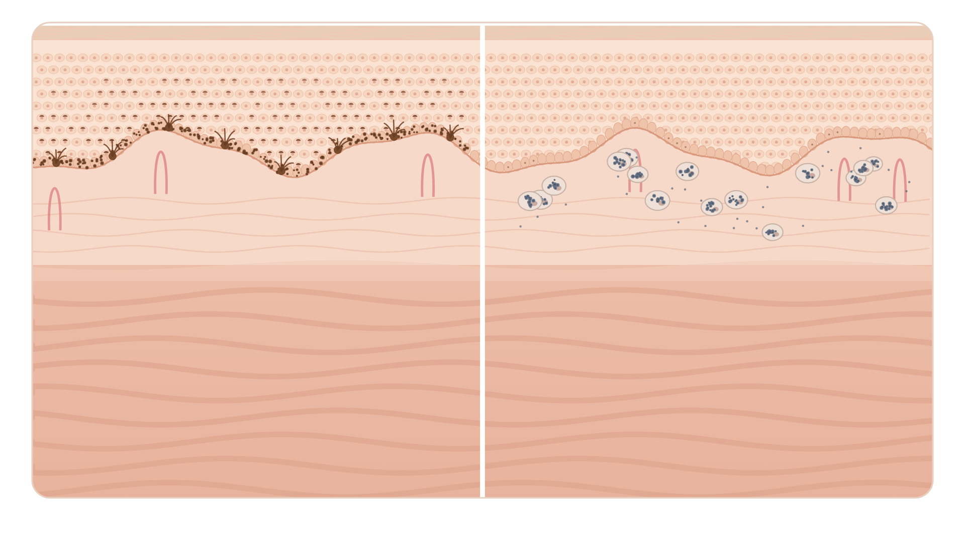
On the surface, this difference often shows up as color. Pigment in the shallow layer tends to look brown, while pigment in the deep layer can look grayish or bluish[2].
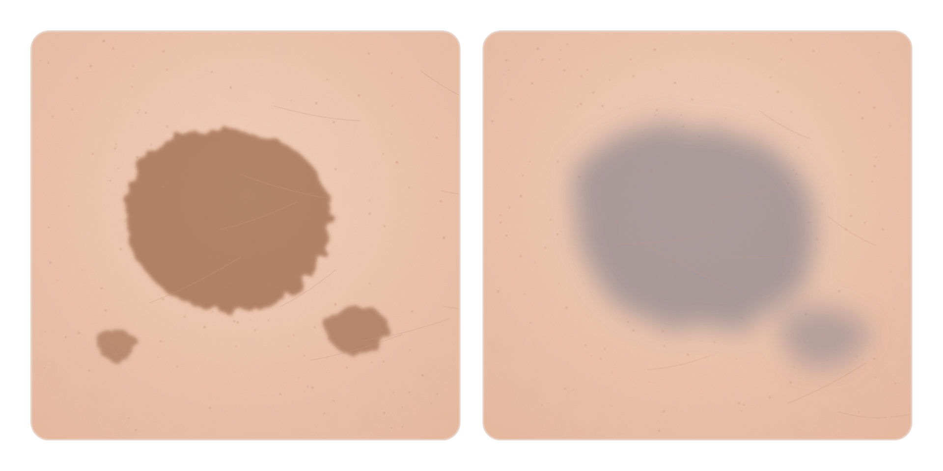
A dermatology society advises that a mark a few shades darker than the surrounding skin usually fades within 6 to 12 months, but it can take years if the pigment is deep. This guidance is written for people with darker skin, so it is better understood as a direction (‘months if shallow, longer if deep’) than as a number that predicts your own timeline[5].
Academic reviews likewise explain that the epidermal type can take months to years without treatment, while the dermal type can last a very long time or remain[2]. That is why I recommend focusing first on “keeping it from getting darker” rather than on “when will it go away.” How you care for the skin during this period can affect how long it lasts.

When is it more likely to happen?
In Korea, there is a randomized study of solar lentigines (sun spots) treated with a 532 nm Q-switched laser (a pigment laser that delivers strong light in a very short pulse). Participants were randomly divided to compare care methods. In the comparison group that applied only a moisturizer, about half of the treated lesions (55.3%) developed PIH. The factors associated with risk in this study were stronger redness at 2 weeks after the procedure and outdoor activity between 1 and 5 p.m. [7]
This figure comes from one specific laser, one specific type of lesion, and 28 participants, so it cannot be carried over as the rate for all laser procedures. Still, it is practical to treat redness that lingers during recovery and midday sun as warning signs.
What can I do at home in the meantime?
1. Sun protection is the foundation
Ultraviolet light can darken pigment that has already formed. A systematic review that pooled many studies also found that broad-spectrum sunscreen with SPF 30 or higher was recommended in nearly all of them[4, 8].
DermNet recommends applying a broad-spectrum SPF 50+ sunscreen every day. So for an area that has already darkened, I recommend a sunscreen that is SPF 50+ and also blocks UVA. [4]
The dermatology society advises using a broad-spectrum sunscreen of SPF 30 or higher, reapplying it every two hours and after sweating or swimming, and wearing a wide-brimmed hat outdoors. It also recommends tinted sunscreen containing iron oxide[5].
Timing matters right after a procedure. As explained in the post on regenerative tape, if the wound is still open or oozing, protect the wound and avoid the sun with shade and a hat instead of applying sunscreen directly. Once new skin has covered it, apply sunscreen consistently.
2. Reducing irritation is part of managing pigment
Because inflammation caused the pigment, it is important not to stir up inflammation again with new irritation. Scrubbing off flakes or putting up with a product that stings can make pigmentation worse. It is better to stop any product that burns or stings when applied[2, 5].
For the same reason, be careful about layering several brightening ingredients at once. If you rush for results and the skin turns red and stings, the attempt to reduce pigment can create new inflammation.
3. Do not expect too much from ‘regeneration creams’
The epidermal growth factor (EGF) ointments introduced in the regenerative tape post showed mixed results across studies. A meta-analysis that statistically combined seven randomized studies of EGF products after laser treatment also found that the proportion who developed PIH was not clearly different from the comparison group, with only a temporary difference in skin pigment measurements at 1 month[9]. Moisturizing to support recovery is necessary, but it is hard to expect any particular regeneration product to prevent pigmentation.
How is it treated at the clinic?
Clinic treatment starts after an examination confirms the cause and the state of the pigment. In broad terms, the order is simple[2].
First, any remaining inflammation is calmed, because pigment keeps being made as long as inflammation continues. Next, a topical medicine that lightens pigment is used together with sun protection. It may sting or dry the skin at first, so the doctor adjusts the amount and frequency while watching how your skin responds[8].
When PIH is likely after a procedure, a short course of a prescription ointment or cream is sometimes used. Studies found less pigmentation, but the doctor decides when to start and how much to use[7, 10]. Do not apply leftover steroid ointment at home on your own. The results come from specific conditions, and misuse can cause other side effects.
Can I just get another laser on the dark area right away? It is better not to rush. The laser can irritate the skin again and make the pigmentation darker. Laser is considered carefully when topical treatment does not lighten it well[2, 4].
When should I see a doctor again?
- When the dark area keeps spreading or getting darker
- When redness, warmth, oozing, or pain persist at the treated spot
- When it looks different in shape or extent from the spot you originally treated
- When it looks bluish-gray and deep pigment is suspected
In these cases, the first step is to check whether inflammation is still present and whether another pigment disorder is also involved. The darker the skin tone, the harder it can be to see remaining inflammation[8].
Let’s return to the original question. Darkening after a laser does not necessarily mean the procedure failed. Blocking the sun, reducing irritation, getting prescription treatment if needed, and giving it time is the most reliable way to let the pigment fade.
Take-home messages
- Darkening after a laser may be post-inflammatory hyperpigmentation, caused by inflammation stimulating pigment cells. It does not necessarily mean the procedure failed.
- Pigment in the shallow layer usually fades over a few months to about a year, but pigment in the deep layer can take years or remain.
- Avoid midday sun with a hat and shade, and once new skin has covered the area, apply broad-spectrum sunscreen consistently (SPF 50+ is recommended for darkened areas).
- Avoid care that creates new inflammation, such as scrubbing, stinging products, and layering brightening ingredients.
- Prescription ointments or creams and additional procedures are timed and chosen by your doctor. Do not apply leftover steroid ointment on your own.
- See a doctor again if the dark area spreads or if redness or pain continues.
References
[1] Kaufman BP, Aman T, Alexis AF. Postinflammatory Hyperpigmentation: Epidemiology, Clinical Presentation, Pathogenesis and Treatment. American Journal of Clinical Dermatology. 2018;19(4):489–503. DOI: 10.1007/s40257-017-0333-6
[2] Davis EC, Callender VD. Postinflammatory hyperpigmentation: a review of the epidemiology, clinical features, and treatment options in skin of color. The Journal of Clinical and Aesthetic Dermatology. 2010;3(7):20–31. PMID: 20725554. PMC2921758
[3] Chan HHL, Manstein D, Yu CS, Shek S, Kono T, Wei WI. The prevalence and risk factors of post-inflammatory hyperpigmentation after fractional resurfacing in Asians. Lasers in Surgery and Medicine. 2007;39(5):381–385. DOI: 10.1002/lsm.20512
[4] Ngan V, Oakley A, DermNet. Postinflammatory hyperpigmentation. Updated 2015. dermnetnz.org
[5] American Academy of Dermatology. How to fade dark spots in darker skin tones. Updated March 2025. www.aad.org
[6] Wolz MJ, Sadler KE, Long CC, Brenner DS, Kim BS, Gereau RW 4th, Kolber BJ. Postinflammatory hyperpigmentation after human cold pain testing. Pain Reports. 2016;1(2):e569. DOI: 10.1097/PR9.0000000000000569 (Source of the Figure 3 photographs, CC BY 4.0)
[7] Kang DH, Choi SM, Lee Y, Kim MS, Lew BL, Kwon SH. Postoperative risk assessment of post-inflammatory hyperpigmentation and the efficacy of delayed prevention following 532 nm Q-switched Nd:YAG laser treatment of solar lentigines: a randomized controlled study. Journal of Dermatological Treatment. 2024;35(1):2398768. DOI: 10.1080/09546634.2024.2398768
[8] Tan MG, Kim WB, Jo CE, Nabieva K, Kirshen C, Ortiz AE. Topical treatment for postinflammatory hyperpigmentation: a systematic review. Journal of Dermatological Treatment. 2022;33(5):2518–2526. DOI: 10.1080/09546634.2021.1981814
[9] Ying J, Zhang Y, Qiu Y, Xiang W. The role of epidermal growth factor-containing topical products on recovery and post-inflammatory hyperpigmentation prevention after laser surgeries: A systematic review and meta-analysis. Journal of Cosmetic Dermatology. 2024;23(2):382–390. DOI: 10.1111/jocd.16007
[10] Cheyasak N, Manuskiatti W, Maneeprasopchoke P, Wanitphakdeedecha R. Topical corticosteroids minimise the risk of postinflammatory hyper-pigmentation after ablative fractional CO2 laser resurfacing in Asians. Acta Dermato-Venereologica. 2015;95(2):201–205. DOI: 10.2340/00015555-1899

Leave a Reply